9082861968 / 8591187089/ 02228893181

Retina

What is Retina?
The retina is the light-sensitive layer of tissue that lines the inside of the eye and sends visual messages through the optic nerve to the brain. The central part of the retina is called macula which is responsible for fine detailed
central vision.

RETINAL DETACHMENT (RD)

What is Retinal Detachment?
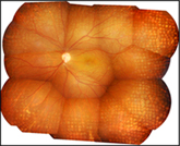
Retinal Detachment (RD) occurs when retina separates from its underlying layers which provide nutritional support to the retina. If not promptly treated, retinal detachment can cause permanent and even complete vision loss.

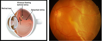
How does retinal detachment occur?
RD may be caused due to tears or holes in the retina (Rhegmatogenous RD), traction on the retina (Tractional RD) or if fluid is built up under the retina (Exudative RD)

What are the risk factors for retinal detachment?
A retinal detachment is also more likely to occur in people who have:

  • High myopia( near sightedness)
  • History of injury to the eye
  • History of retinal detachment in the other eye
  • Family history of retinal detachment
  • History of cataract surgery
  • Inflammatory eye disease

What are the symptoms of retinal detachment?

  • Sudden onset or increase in floaters
  • Flashes of lights
  • Gray curtain or veil moving across your field of vision
  • Sudden painless loss of vision

What are floaters and flashes of light?
Floaters are small, dark, shadowy shapes that can look like spots, thread-like strands, cob webs or squiggly lines that move in your field of vision. Flashes look like flashing lights or lightning streaks in the peripheral vision even though no light is actually flashing. Floaters are very common and is usually not a sign of a serious condition. However, if floaters and/or flashes begin or increase suddenly, it may indicate a serious problem, and hence needs immediate medical attention.


What is the treatment of retinal detachment?
RD can be treated in any of the following ways:

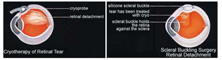
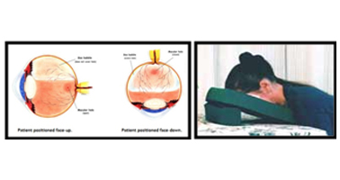
Pneumatic retinopexy involves injection of a gas bubble into the vitreous (the jelly-like substance in the eye cavity) enabling the gas bubble to push the retinal tear back against the wall of the eye and close the tear. Laser or cryopexy (freezing) is used to secure the retina to the eye wall around the retinal tear. .

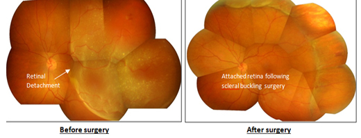
Scleral buckle surgery involves suturing a silicone “buckle” to the eye that indents the wall of the eye to support the retinal breaks allowing the retina to reattach.Cryopexy (freezing) treatment is usually used to seal the retinal tears.
The silicone buckle is usually left on the eye permanently. Someone looking at you cannot see the buckle, as it is stitched to the sclera somewhere towards the back of the eye.

At Kumta Eye and Retina clinic, we perform highly specialized minimally invasive buckling technique called Minimal Scleral Buckling.


Vitrectomy involves removing the vitreous gel and filling the eye with gas or silicone oil bubble. Gas or silicone oil placed in the vitreous cavity helps to hold the retina in place.The holes and tears are sealed with laser, and fluid under the retina is drained.
If silicone oil has been used, it requires a second surgery to remove the oil, at a later date. Gas bubble doesnot require to be removed and usually gets absorbed within 4 weeks.

At Kumta Eye and Retina clinic, we perform MIVS (Microincisional Vitrectomy Surgery) using very tiny self sealing incisions which do not require sutures and hence recovery is very quick, healing time is much faster and postoperative discomfort, pain, and redness is minimal as compared to conventional vitrectomy. This is performed with the most advanced high speed vitrectomy machine (Constellation). (Scroll down for more details)


What is the success rate of retinal detachment surgery?
The anatomical success rate(chances of retina getting attached) is about 90% .There is a small risk (about 10%) of redetachment of the retina after surgery, in which case, a repeat surgery to reattach the retina may be required. This usually occurs if there is a scar tissue formation over the retinal surface called proliferative vitreoretinopathy (PVR) which causes the retina to redetach.
The functional success rate (visual outcome) depends on factors like duration and type of detachment, age of the patient. Although the retina gets settled well after surgery, vision remains compromised in these cases, since there is permanent damage to the vision cells of the retina whenever the retina gets detached.

RETINAL VEIN OCCLUSION (RVO)

What is retinal vein Occlusion (RVO)?
RVO occurs when there is a blockage in the retinal vein (which drains blood away from the retina) causing the walls of the vein to leak blood (haemorrhage) and fluid into the retina. Fluid accumulation under the macula causes sudden painless blurring of vision. If RVO is not treated, it can sometimes lead to complete loss of vision.,

What are the types of RVOs?
RVOscan be mainly of two types:

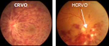
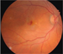
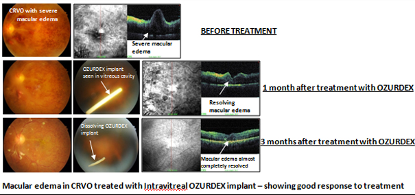
Central Retinal Vein Occlusion (CRVO) –occurs when the main (central) retinal vein gets blocked, thus leading to involvement of the entire retina. Less commonly, Hemi central retinal vein occlusion (HCRVO) may occur when there is blockage of a vein draining the superior or inferior half of the retina.


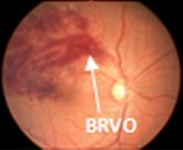
Branch Retinal Vein Occlusion (BRVO)occurs when there is a blockage within a branch of the retinal vein leading to hemorrhage along the distribution of a small vessel of the retina.

What are the types of CRVO?
Non ischemic CRVO – it is a is a milder form of the disease where the blood supply of the retina is not affected Ischemic CRVO – it is a more serious form where the blood supply of the retina is lost, hence can lead to the development of significant complications,vision loss and rarely loss of eye.

What are the symptoms of RVO?
Patient with RVO may complain of sudden painless loss of vision in the affected eye. There may be minimal or no symptoms if macula is not affected.

What are the risk factors for RVOs?
High blood pressure, Diabetes, High cholesterol, elevatedHomocysteine levels, Ocular hypertension & Glaucoma, Fasting especially water deprivation

What is the treatment for RVO?
There is no treatment proven to reopen occluded retinal veins. Strict control of all risk factors is very important.

Intravitreal Anti-VEGF injections ( Lucentis /Avastin)–to reduce macular edema and improve vision. It may sometimes be combined with a steroid injection.

Intravitreal steroid injections (Triamcilnolone/ Ozurdex implant) – to reduce macular edema and improve vision. Can be combined with Anti VEGF injections.

Laser Photocoagulation-Laser treatment is applied to whole peripheral retina (PRP in ischemic CRVO) or affected retinal area in BRVO to decrease the risk of further complications. Laser does not improve sight.

Vitrectomy – May be required in cases of non resolving vitreous haemorrhage due to RVO.


AGE RELATED MACULAR DEGENERATION (AMD)

What is Age related macular degeneration (AMD)?
AMD is a disease that is associated with aging that gradually destroys the macula thus affecting central vision in the affected eye.

What are the types of AMD?
AMD occurs in two forms:


Dry AMD -Occurs when the light sensitive cells in the macula slowly break down gradually blurring central vision.


Wet AMD - Occurs when new abnormal blood vessels which are very fragile start growing beneath the macula which may often leak blood and/or fluid causing rapid loss of central vision.

What are the Symptoms of AMD?
Blurred central vision, distorted vision, difficulty in recognizing faces and reading


What is the treatment for AMD?
There are two main treatment options for wet AMD:

  • Intravitreal Anti-VEGF injections (Lucentis (FDA approved)/ Avastin(non FDA approved)) to prevent the growth of new blood vessels.
  • Laser treatment to destroy abnormal blood vessels in the eye.
  • Photodynamic therapy (PDT) with Visudyne - PDT uses a light sensitive dye – Visudyne which when injected, accumulates in abnormal blood vessels in the eye and a cold laser is used to activate the drug which then seals the abnormal vessels without damaging the overlying sensory retina.

DIABETIC RETINOPATHY (DR)

What is Diabetic Retinopathy?
Diabetic Retinopathy (DR) is a progressive damage to the tiny retinal blood vessels caused by complications of long-standing diabetes, which if not treated at an appropriate stage can eventually lead to complete loss of vision

What are the symptoms of Diabetic retinopathy?
In DR, patient may be asymptomatic (no vision complaint) unless it reaches an advanced stage or macula is affected. Hence every diabetic should get a comprehensive dilated eye examination done once in a year for early detection of DR.
In Diabetic macular edema, patient may have blurred vision, difficulty in reading, and recognizing faces.
In PDR with vitreous haemorrhage, patient may complain of sudden drop in vision with associated floaters.

What are the stages of Diabetic Retinopathy?
DR can be classified into two stages:

Non proliferative diabetic retinopathy (NPDR)– is an early form of the disease where patient may usually not have any vision complaints. It is classified as mild, moderate and severe NPDR depending on the amount of retinal damage.


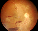
Proliferative diabetic retinopathy (PDR) – is a more advanced form of the disease where new abnormal blood vessels grow on retinal surface (in response to the loss of blood supply caused by closure of normal blood vessels) which can bleed anytime (vitreous haemorrhage) or cause pull on the retina (tractional retinal detachment) leading to sudden loss of vision. Neovascular glaucoma (NVG) may occur in end stages which may lead to a painful blind eye.


What is Diabetic macular edema (DME)?
DME is swelling or thickening of the macula due to fluid leakage from damaged blood vessels within the macula due to diabetes causing blurring of vision. DME can occur at any stage of DR.

How is Diabetic Retinopathytreated?
In the early stages of NPDR, treatment other than regular monitoring is not required. Strict control of diabetes, blood pressure and cholesterol is very important to control progression of DR.

  • Laser Photocoagulation- In severe NPDR or PDR, laser treatment is applied to peripheral retina (PRP) to decrease the risk of complete vision loss. Laser does not improve sight.
    At Kumta Eye and Retina Clinic, we use the latest laser technology including MULTISPOT PATTERN LASER & MICROPULSE which have very less side effects & virtually painless (scroll down for more details)
  • Intravitreal Anti-VEGF injections ( Lucentis /Avastin)– In cases of vitreous haemorrhage due to PDR, anti VEGF injections cause new abnormal blood vessels to shrink and hence stop further bleeding.
  • Vitrectomy (with or without silicone oil injection)– In advanced stages of PDR, surgery may be indicated.

    What is the treatment for DME?
    DME if mild can be treated directly with laser. If edema is more, then intravitreal injections are first required to reduce the swelling and then laser is done to seal leaking blood vessels.The injections can be of 2 types:
  • Intravitreal Anti-VEGF injections ( Lucentis /Avastin) - It may sometimes be combined with a steroid injection.
  • Intravitreal steroid injections (Triamcilnolone/ Ozurdex implant) - Can be combined with Anti VEGF injections.

ENDOPHTHALMITIS

What is Endophthalmitis?
It refers to a serious and vision threatening infection inside the eye that requires urgent treatment. It is the most dreaded complication after any eye surgery. It may be bacterial or fungal.If not treated promptly, it may lead to complete loss of vision.

What are the types of Endophthalmitis?
Exogenous Endophthalmitis is more commonly seen. It is caused by penetration of the eyeball by infectious organism from outside which may be due to:
  • Eye surgery
  • Trauma
  • Intravitreal Injections
  • Corneal infection
Endogenous Endophthalmitis is caused by organism which reaches the eye through the bloodstreamfrom a distant source of infection in the body. It is relaltively rare.

What are the symptoms of endophthalmitis?
Sudden loss of vision,pain, redness, intolerance to light and floaters.

What is the treatment of endophthalmitis?
Prompt treatment is necessary for better visual outcome. A sample of the fluid from inside the eye (aqueous/ vitreous humor) is aspirated and sent for testing to help determine the causative organism and concurrent injections of intravitreal antibiotics are given. In more severe forms, urgent vitrectomy(surgery) may be required to remove the infection.

The outcome largely depends on what organism was involved in the infection.

VITREORETINAL INTERFACE DISEASES

What are Vitreoretinal Interface Diseases?
The vitreous gel is strongly attached to the retina (at macula) since birth. This gel starts liquefying and separating from the retina with age. Changes in vitreous gel may cause pull on the retinal surface and macular distortion. Some of the commonly seen vitreoretinal interface diseases are:

  1. 1. Vitreomacular traction syndrome (VMTS)
  2. 2. Epiretinal membrane (ERM) or Macular pucker
  3. 3. Full thickness macular hole (FTMH)

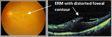
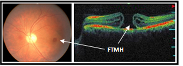
What are the symptoms of Vitreoretinal diseases?
Since this is a macular disease, central vision is mainly affected - blurred central vision with difficulty reading and recognizing faces, distorted vision, gray spot in central vision (central scotoma)


What is Vitreomacular traction syndrome (VMTS)?
VMTS occurs when, vitreous separates partially from the retina with persistent attachment to the macula (centre of the retina), causing pull on the macula thus leading to swelling in the macula.
How is VMTS treated?
The treatment of VMTS is vitrectomy surgery to release all adhesions of the gel to the macula, and to peel away any residual membranes on the surface of the macula that may also be contributing to distortion of the vision. Sometimes the eye is filled with a gas at the end of surgery to help with healing. If used, the gas slowly disappears over days to weeks.

What is Epiretinal membrane (ERM) or Macular pucker?
ERMis a film of tissue which grows on the surface of the macula, which may sometimes cause distortion and/or swelling in the macula.


What is the treatment of ERM?
If there are no or minimal visual symptoms, ERM can be observed and does not require surgery. ERM needs to be removed surgically, if it causessignificant swelling in the macula and significant drop in vision. ERM surgery involves vitrectomy (removing the vitreous gel) and gently peeling of the membrane from the surface of the macula using very fine instruments.Sometimes the eye is filled with a gas at the end of surgery to help with healing. If used, the gas slowly disappears over days to weeks.


What is Full Thickness Macular Hole (FTMH)?
It is a defect (hole) in the macula thus affecting the central vision.
What are the causes of FTMH?
Although, most of the macular holes are idiopathic (unknown cause), there are some associations with injury and high myopia (short sightedness).


What is the treatment of FTMH?
Almost all macular holes require surgery. The prognosis of the surgery depends on the size and duration of the hole. Smaller and recently developed holes have greater anatomical (hole closure)and functional (visualimprovement) success rate as compared to larger and long standing holes. Macular hole surgery involves doing vitrectomy (removing the vitreous gel) and filling the eye with gas which acts as a tamponade allowing the hole to close.In order to maximize the effect of the surgery, the patient is usually required to remain in a face down position for one week postoperatively to allow the gas bubble to exert pressure against the edges of the hole thus allowing the hole to close. The bubble will gradually be reabsorbed as the vitreous cavity refills with a naturally produced eye fluid.


The other retinal conditions treated at Kumta Eye & Retina Clinic are
Retinopathy Of Prematurity (ROP), Ocular trauma & Management of cataract surgery complications like dislocated nucleus, dislocated Intraocular lens (IOL), and endophthalmlitis.


Facilities Available

Non-mydriatic digital fundus photography, Fundus Fluorescein Angiography (FFA) from Kowa (Japan)
  • FFA refers to photographing the retinal vasculature following intravenous injection of fluorescein dye.
  • It helps in confirming diagnosis, quantifying severity of the disease and decide mode of treatment.
  • The non mydriatic function in the camera enables imaging retina even through small pupil.

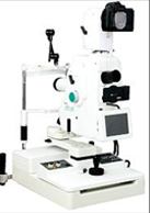
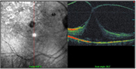
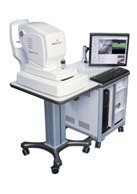
Spectral Optical coherence tomography (3D OCT/SLO) from OPKO (USA)

  • It is a non contact and rapid imaging of macula using light
  • It provides high-resolution, cross-sectional images of the macula
  • Can also be used to examine retinal nerve fiber layer and the optic nerve head
  • Used for diagnosis, documentation, follow up & assessment of response to therapy of various macular diseases :Diabetic macular edema; Macular edema due to retinal veinocclusions; Choroidal neovascular membranes & ARMD; Vitreoretinal interface diseases (Macular hole, pucker, VMTS); Uveitis; unexplained visual loss
  • It can pick up subtle macular abnormalities which can be missed on clinical examination.

Multispot Pattern Laserfrom Iridex (USA)
Laser treatment involves using an intense beam of light (laser) that can be precisely focused on the retinal areas to be treated.
It is widely used for treatment of Diabetic retinopathy and other retinal vascular disorders. It is also used to seal retinal tears or holes in patients as preventive treatment of retinal detachment.
As compared to conventional laser, multispot laser is extremely fast, require shorter sessions, causes less damage to surrounding tissues hence lesser visual defects and causes less pain hence well tolerated by patients,br>

Advanced Vitreo-Retinal Surgical Facilities with Constellation combined vitrectomy system from Alcon (USA), Leica M501 zoom operating microscope with XY & Camera (Swiss), BIOM & sutureless MIVS (Microincisional Vitrectomy Surgery) technology

Constellation is the best vitrectomy machine available with MIVStechnology

It is a latest sutureless vitrectomy surgery with very tiny incisions as compared to 20 gauge procedure.

This system has combined capabilities to perform advanced sutureless vitreo retinal surgery, Ozil - torsional phacoemulsification for cataract surgery and Retinal Lasers with endo-laser facility for diabetic retinopathy and managing complicated retinal detachment cases.

Sutureless vitrectomy
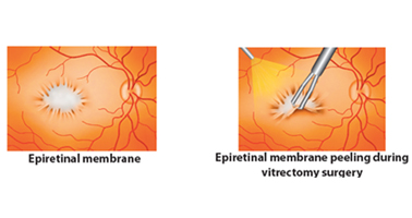
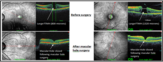
What isMicroincisional Vitrectomy Surgery(MIVS)?

Vitrectomy is a surgical procedure that involves removal of vitreous gel from the eye by making incisions in the sclera (the white part of our eye) to gain access into the vitreous cavity and the retina. In conventional vitrectomy,these incisions are relatively large and sutures are required to close these incisions.
In Microincisional Vitrectomy Surgery,the incisionis made with specialized instruments and is much smaller than in conventional vitrectomy, and therefore they close by themselves and donot require sutures.MIVS is done with a specialized vitrectomy system called Constellation.There are 3 techniques available in MIVS: 23 Gauge, 25 Gauge and 27 Gauge Vitrectomy

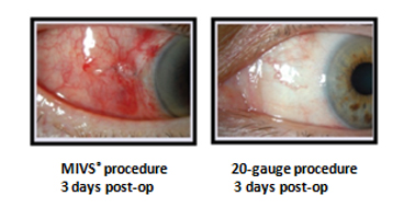
What are the advantages of MIVS?
Since it is sutureless, foreignbody sensation related to sutures is not there. Since size of the incisions in MIVS is very tiny, healing time is much faster and postoperative discomfort, pain, and redness is minimal as compared to conventional vitrectomy.


What are the limitations of MIVS?
Although MIVScan be done in most of the retinal diseases, there are some conditions where conventional vitrectomy would be required. The retinal surgeon would be the best judge to decide if MIVS would bepossible in your case or not.


Intravitreal Anti VEGF injections (Lucentis/ Avastin)
Lucentis (Ranibizumab) (FDA approved) and Avastin (Bevacizumab) (non FDA approved) injections are used to treat wet age related macular degeneration,diabetic macular edema, macular edema due to retinal vein occlusions, vitreous hemorrhage, retinopathy of prematurity, and Choroidal neovascularisation membrane due to other causes.


Intravitreal Ozurdex implant
It is a rod shaped steroid implant placed inside the vitreous cavity which is slowly released into the eye. It is used to treat macular edema due to retinal vein occlusion, diabetic macular edema.


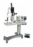
Photodynamic therapy (PDT)with Visudyne
PDT uses a light sensitive dye – Visudyne which when injected, accumulates in abnormal blood vessels in the eye and a cold laser is used to activate the drug which then seals the abnormal vessels without damaging the overlying sensory retina. It is used in treatment of wet age related macular degeneration, Chronic Central serous retinopathy